Time resolved laser fluorescence spectroscopy (TRLFS) is the analytical technique which observes de-excitation processes of excited fluorophore created by short-pulsed laser. TRLFS data of a fluorescent metal ion includes
- shape of emission spectrum
- its temporal decay
These information reflects
- the number of allowed transitions and their probabilities
- competitive non-fluorescent de-excitation pathways
Thus, the spectral shape will change when a fluorescence metal coordinates with a ligand and changes its energy levels and/or associated transition probabilities due to variation of the symmetry around it. Similarly, fluorescence decay lifetime will vary when a ligand around the central metal ion is displaced by another ligand, as de-excitation pathways change. We can use these changes in TRLFS data to probe reactions with a fluorescent metal ion and estimate its structural information. As good approximation,
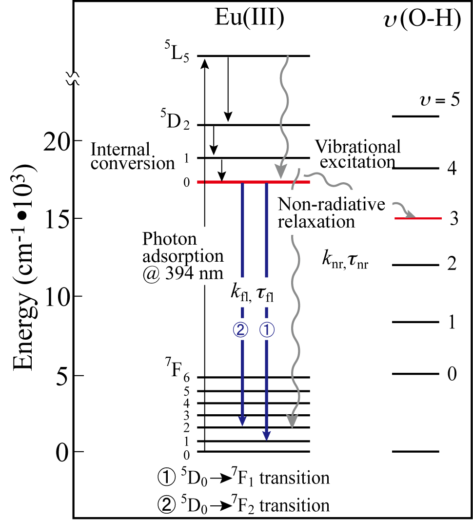
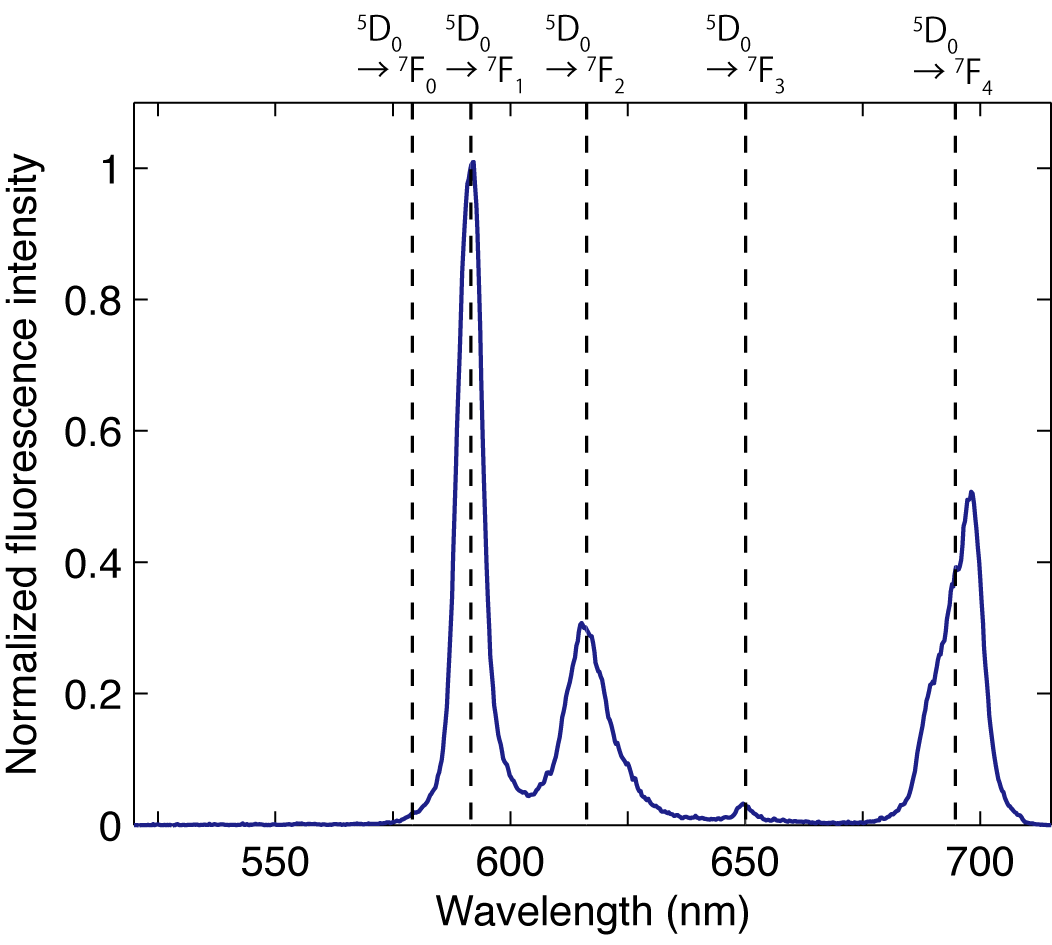
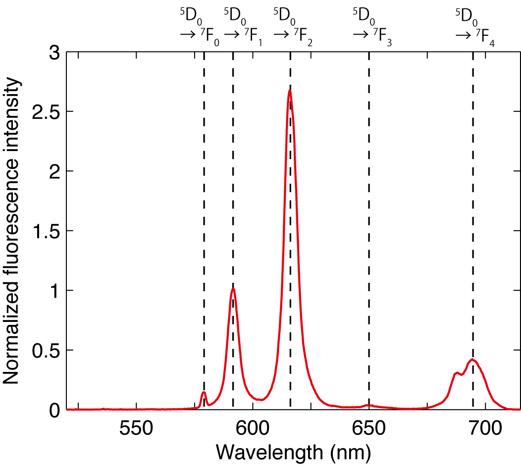
For instance, Eu3+, a chemical homologue of trivalent actinides such as Am3+ and Cm3+, has the energy diagram as in Figure 1 and the emission spectrum as in Figure 2. Its decay lifetime is around 110 μsec. Three major peaks in Figure 2 corresponds to the transitions from the 5D0 states to the 7F1,7F2,and 7F4 states. Upon complexation with acetate ligand, the spectrum changes in Figure 3. Particularly, the peak around 620 nm due to so-called hyper-sensitive transition, 5D0 → 7F2, is significantly enhanced. An overtone of OH vibration of water molecule provides an efficient pathway for excited Eu3+; the decay lifetime is increased as a part of hydration water molecules is replaced with acetate. As a good approximation, the reciprocal of fluorescence lifetime of Eu3+ is known to be proportional with the number of remaining water molecules in its first coordination sphere.
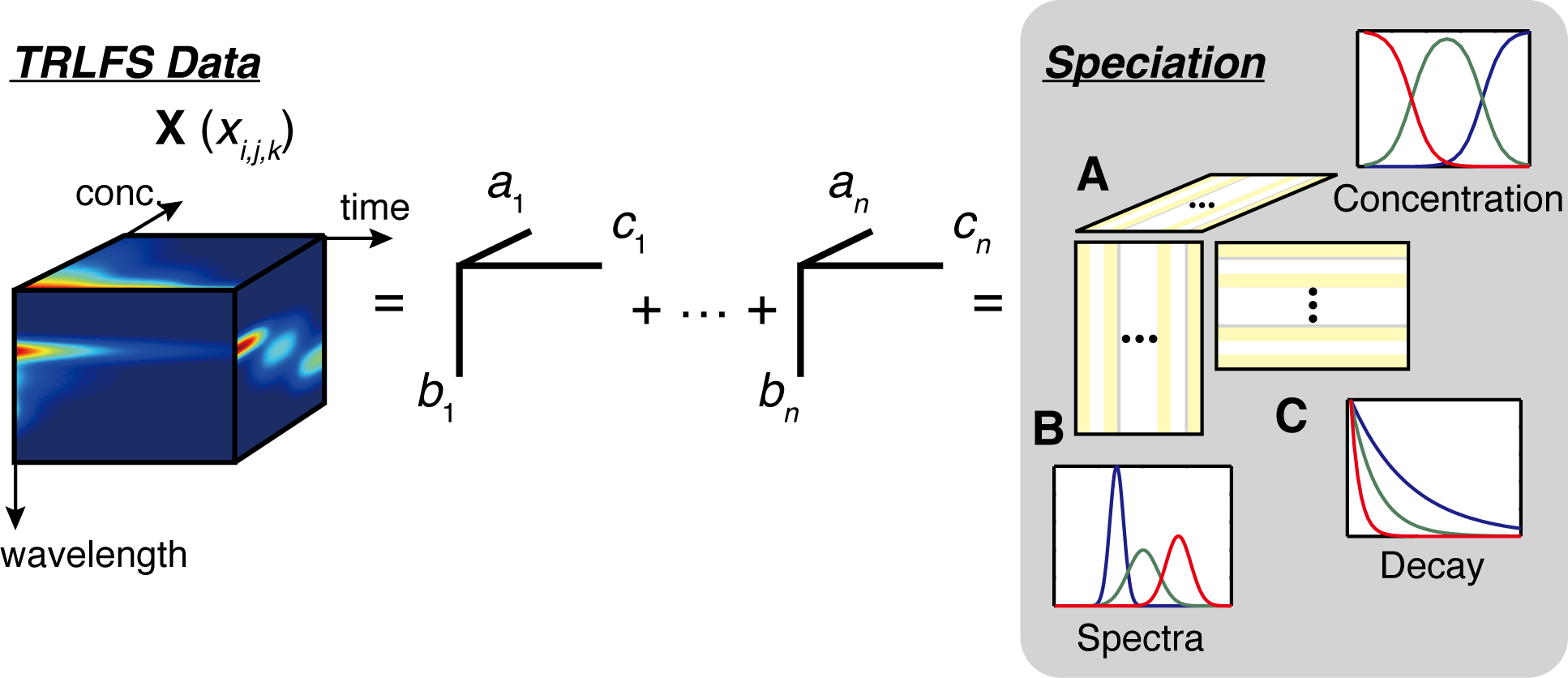
TLRFS is a very selective speciation technique, as it provides multivariate data defined with emission wavelength and decay time. Moreover, its quantitative quality is also good, as the fluorescence intensity is proportional to the concentration of an underlying species of a fluorescent metal ion. In our laboratory, we are investigating complexation of fluorescent metal ions with small ligands, their adsorption on mineral surfaces, and binding to natural organic matters, using a multi-mode factor analysis called parallel factor analysis (PARAFAC, Figure 4).
Sasaki, T*, Ueda, K., Saito, T., Aoyagi, N., Kobayashi, T., Takagi, I., Kimura, T., Tachi, Y., “Sorption of Eu3+ on Na-montmorillonite studied by time-resolved laser fluorescence spectroscopy and surface complexation modeling”, J. Nucl. Sci. Technol., in press (2015).
Saito, T.*, Aoyagi, N., Kimura, T., “Time-resolved laser-induced fluorescence spectroscopy combined with parallel factor analysis: a robust speciation technique for UO22+”, J. Radioanal. Nucl. Chem. 303, 1129-1132 (2015).
青柳登,斉藤拓巳,木村貴海,「レーザー分光を用いたアクチノイドの状態分析」,ぶんせき9, 536-542, 2013.
Lukman, S., Saito, T.*, Aoyagi, N., Kimura, T. , Nagasaki, S., “Speciation of Eu3+ Bound to Humic Substances by Time-Resolved Laser Fluorescence Spectroscopy (TRLFS) and Parallel Factor Analysis (PARAFAC)”, Geochim. Cosmochim. Acta 88, 199-215 (2012).
Ishida, K., Saito, T.*, Aoyagi, N., Kimura, T., Nagaishi, R., Nagasaki, S., Tanaka, S., “Surface Speciation of Eu3+ Adsorbed on Kaolinite by Time-Resolved Laser Fluorescence Spectroscopy (TRLFS) and Parallel Factor Analysis (PARAFAC)”, J. Colloid Interface Sci.374, 258-266 (2012).
Collins, R. N.*, Saito, T., Aoyagi, N., Payne, T. E., Kimura, T., Waite, T. D., “Application of Time-Resolved Laser Fluorescence Spectroscopy to the Environmental Biogeochemistry of Actinides”,J. Environ. Qual. 40, 731-741 (2011).
Saito, T.*, Sao, H., Ishida, K., Aoyagi, N., Kimura, T., Nagasaki, S., Tanaka, S., “Application of Parallel Factor Analysis for Time-Resolved Laser Fluorescence Spectroscopy: Implication for Metal Speciation Study”, Environ. Sci. Technol. 44, 5055-5060 (2010).
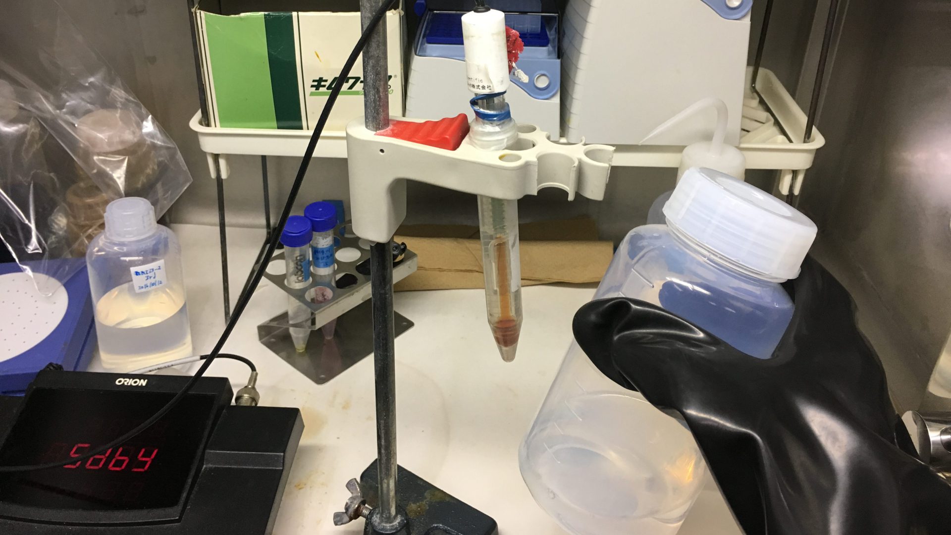
Overview of our TRLFS set-ups
This research is perfomred under collaboration with the research group for radiochemistry, Japan Atomic Energy Agency.
Laser sources
- Ti:sapphire femto-sec laser + regenerative amplifier (~800 or ~400 nm, Spectraphysics Tsunami + Spitfire)
- Nd:YAG nano-sec laser + OPO(variable wavelength, Spetracphysics社 Quanta-Ray + versaScan, uvScan)
- Nd:YAG nano-sec laser (1064, 532, 355, 266 nm, Quantel Brilliant)
- Nd:YAG nano-sec laser(532 nm, LOTIS TII)
Spectrometer and detectors
- Andor Shamrock spectrometer+ iStar ICCD camera
- Acton spectrometer + Roper PI-MAX3 ICCD camera
- Chromex spectrometer + Hamamatsu Photonics streak camera system (C7700)
Microscopy
- Scanning near field optical microscopy (SNOM, JASCO)
Accesaries
- UV-Vis, IR cryostat for spectroscopy (Unisoku, CoolSpeK)
- Cryogen-free cryostat for spectroscopy (Oxford Opticool)
Applications
- Femto-sec TRLFS
- Nano-sec TRLFS
- Ramman scattering
- Scanning near field optical microscopy